Understanding the anatomy of a leaf is a handy piece of knowledge to have. Knowing how all the tree parts work together will help you become a better arborist.
In this article, I will cover the anatomy of leaves. I will list each part of the leaf and describe its function. I hope by the end of the article you’ve learned something new.
Introduction
When learning the anatomy of a leaf, we must understand there are two different types of leaves. These are:
- Angiosperm Leaves
- Gymnosperm Leaves
From here, botanists have broken down leaves even further. Breaking down leaves by shapes, sizes, etc, can help with tree identification. In this article, I won’t be talking about these differences. I’ll just cover the anatomy.
Angiosperm and gymnosperm leaves aren’t totally different. There are some similarities they both share. One major similarity is the xylem and phloem.
Xylem and Phloem
The xylem and phloem make up a tree’s vascular system. These veins will help transport water and minerals into the leaf. They will also help carbohydrates out of the leaf to the rest of the tree.
The xylem will transport water and minerals into the leaf. The phloem will move carbohydrates within and out of the leaf.
Angiosperm Leaf Anatomy
Angiosperm leaves tend to be large and broad. This will help the leaves absorb as much sunlight as possible.

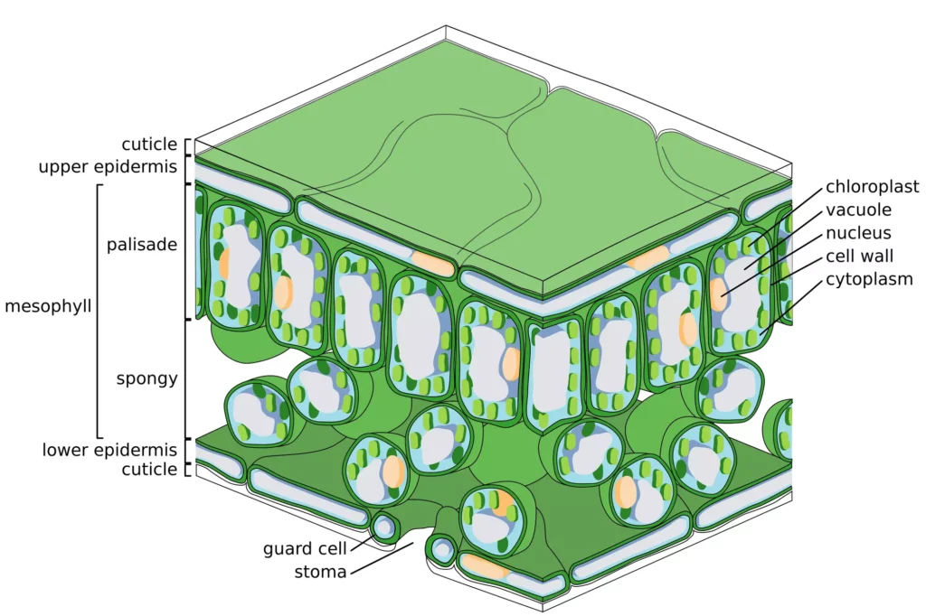
Cuticle
The cuticle is a waxy coating on the outside of the leaf. This layer is waterproof and prevents water from getting into the leaf and escaping the leaf. The substance that gives the cuticle its waxy texture is cutin.
The thickness of the cuticle will vary depending on the leaf’s environment. For example, shade leaves will have a thinner cuticle layer than sun leaves.
Shade leaves don’t suffer as much water loss as shade leaves do. So, the cuticle on shade leaves is thinner. Sun leaves face intense sunlight, so they need a thick cuticle to prevent water loss.
To learn more about sun and shade leaves, check out this article.
Upper Epidermis
The epidermis is a layer of cells surrounding the leaf. The upper epidermis is on the top side of the leaf. In contrast, the lower epidermis is on the underside.
The epidermis is transparent, allowing in as much sunlight as possible. The sunlight will travel through the epidermis into the leaf, where the photosynthetic material is located.
Mesophyll
The mesophyll is the inside section of a leaf. This section contains mostly parenchyma cells. Parenchyma cells are cells that carry out essential functions.
The mesophyll is broken down into two sections:
- The palisade mesophyll
- The spongy mesophyll
Palisade Mesophyll
The palisade mesophyll is column-like cells that are essential to photosynthesis. Inside these cells are chloroplasts. The chloroplasts contain chlorophyll which absorbs sunlight. Photosynthesis happens in the chloroplasts.
Sun leaves will have extra layers of palisade mesophyll. This is to help sun leaves deal with all the light energy they receive. If a leaf can’t use all the light it receives, the light will damage the leaf. By having more photosynthetic material, these leaves can use more light.
Some trees have palisade mesophyll in the upper and lower levels of the leaf. These trees can photosynthesize light on both sides of the leaf. Also, these leaves can reduce heat load and water loss.
Spongy Mesophyll
A spongy mesophyll is a group of loosely packed cells. These cells also have an irregular shape, allowing plenty of airflow through this layer.
This section of the leaf is where gas exchange happens. A thin layer of water covers these cells. Gas will dissolve in this water.
When the leaf is photosynthesising, carbon dioxide can diffuse into these cells, and oxygen will diffuse out.
Stoma (Stomata Plural)
Think of the stomata as tiny pores on the underside of the leaf. These little pores allow carbon dioxide to enter the leaf. The stomata will also allow oxygen and water to exit the leaf.
Most angiosperm trees will have stomata on the underside of the leaf only. This will prevent water loss in direct sunlight. However, some trees may have stomata on both sides of the leaf. These trees will do better in high sunlight conditions.
Guard Cells
Two guard cells surround each stoma. These guard cells control the opening and closing of the stoma.
When conditions are good, the guard cells will fill up with water and allow the stomata to be open. The tree will be able to absorb carbon dioxide to assist in photosynthesis.
However, the guard cells will keep the stomata closed when water is scarce. When the stomata are closed, water is not able to escape. So, the tree will retain as much water as possible to survive.
Photosynthesis will slow down if the stomata are closed. This is because carbon dioxide will not be able to enter the leaf. So, if a tree has its stomata closed for too long, the tree will die.

Gymnosperm Leaf Anatomy
Gymnosperm leaves can come in various shapes and sizes. The traditional-looking leaves will have a very similar structure to angiosperm leaves. However, needles like those on pine trees come under gymnosperm leaves. These leaves will be quite different from angiosperms.
The diagram below is that of a pine needle. Using a pine needle, I can highlight some of the different anatomy found in gymnosperm leaves.
There are still some similarities:
- Epidermis
- Cuticle
- Phloem
- Xylem
- Stoma
- Guard Cell
I won’t cover these topics in this section. They have the same function as mentioned earlier in this article.
Resin Duct
Many Gymnosperm species have resin ducts on their leaf. These resin ducts allow the transportation of resin throughout the tree.
The resin is a sticky substance that oozes out of trees in response to injury. Resin will help protect the tree from insects and microorganisms. Resin can stop these threats by:
- Being toxic to pathogens
- Physically blocking insects
Resin is not to be confused with sap. The sap is a watery, sugary substance. Whereas resin is thick and sticky.
Transfusion Tissue
Let’s look to Merriam-Webster for a definition of transfusion tissue.
Tissue that is found characteristically around the vascular bundles of gymnosperm leaves and consists of both living cells like those of parenchyma with walls that are not lignified and thin-walled but lignified tracheids with bordered pits. Source: Meriam-Webster
So, the transfusion tissue surrounds the xylem and phloem. The cells that make up transfusion tissue help control the movement of substances. These substances will move into and out of the xylem and phloem.
Endodermis
The endodermis surrounds the transfusion tissue. This part of the leaf will help regulate material flow in and out of the vascular structures.
Chlorenchyma
Chlorenchyma is the name given to the cells containing chlorophyll in some species of gymnosperms. Species with chlorenchyma tend to have needles rather than traditional leaves. So, an example of a tree with chlorenchyma is a pine tree.
Gymnosperms with traditional leaves will have palisade mesophyll, just like most angiosperms.
Hypodermal Sclerenchyma
The hypodermal sclerenchyma layer sits just underneath the epidermis. This layer is what makes pine needles so rigid.
The cells in this layer are rich in lignin. Lignin is a rich organic material that is important for mechanical support. Hence, pine needles are so stiff and upright. Lignin is vital for mechanical support; it also aids in water transport and helps a tree resist stressors.
